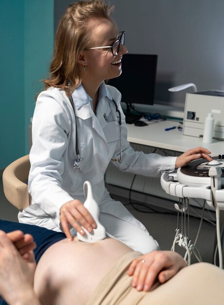

2D Echocardiogram (2D Echo) is a non-invasive ultrasound technique that uses high-frequency sound waves to create real-time, two-dimensional images of the heart. It’s one of the most commonly used tests to assess the structure and function of the heart. The procedure helps doctors evaluate the heart’s chambers, valves, and major blood vessels, and it is crucial for diagnosing various heart conditions.
2D echocardiogram is a crucial tool in diagnosing and managing heart conditions. It provides clear, real-time images of the heart’s structure and function, helping doctors make informed decisions about treatment and care.
– Assessing Heart Function: To evaluate how well the heart pumps blood, detect heart failure, or measure ejection fraction.
– Diagnosing Heart Valve Problems: To check for conditions like valve stenosis, regurgitation, or prolapse.
– Investigating Heart Disease: To detect structural abnormalities such as holes in the heart, thickened walls, or heart muscle disease.
– Monitoring Cardiac Conditions: For patients with a history of heart disease or those undergoing treatment for heart conditions, a 2D echo is used to monitor progress or the effectiveness of interventions.
– Evaluating Blood Flow: It can be used alongside Doppler imaging to assess blood flow through the heart’s chambers and valves.
– No Radiation: Unlike other imaging techniques such as X-rays or CT scans, 2D echo uses sound waves, so there’s no exposure to radiation, making it safe for repeated use, especially in sensitive populations like children or pregnant women.


© Copyright 2025 - Tech Scan Cure Diagnostic
© 2025 Designed By Bazaar Digital : A creative Agency